Secondary Antibodies for Super Resolution Microscopy Jackson ImmunoResearch

Secondary antibodies conjugated with Alexa Fluor® 488 / 594 / 647, FITC and Cy5 for STED and STORM.

JIR Antibody Dye Conjugates for use in SuperResolution (SR) Microscopy

Super-resolution microscopy (SRM) encompasses any optical technique which circumvents the resolution limits of light diffraction found in conventional light microscopy. SRM techniques allow cellular structures to be resolved to the sub-organelle level, enabling information about the 3D structure of cellular components to be determined, and single molecule colocalization to be observed.

Each SRM technique has its own requirements for probe selection. Jackson ImmunoResearch offers a wide selection of labeled secondary antibodies with dyes known to be robust in SRM methods. It should be noted that the field of SRM changes rapidly and guidance for each of the techniques is beyond the scope of this catalog. It is therefore recommended that information from specialized technical literature is sought to aid fluorophore selection.

The following sections briefly outline the principles of some of the most popular SRM techniques:

Stimulated Emission Depletion (STED) microscopy

Diffraction of light by a microscope lens causes light from a single point to appear over a larger area, known as a point spread function or PSF (see Figure 38).
Stimulated emission depletion (STED) microscopy produces super-resolution images by confining the fluorescing region (PSF) of a sample. STED microscopes use two overlapping lasers, the first of which excites the fluorophores as per conventional microscopy (Figure 39A). The second laser, called a depletion laser (STED laser), excites a “donut” of light, with a very small (~30 nm) zero intensity (unexcited) point at its center. This second laser essentially “switches off” the fluorescence generated by the first laser except at the center of the donut, thereby reducing the excited fluorescent molecules to those at the zero point. This effectively reduces the PSF to produce a very small focused region of single molecule fluorescence. Without overlapping interference patterns, high-resolution images can be obtained (Figure 39D). Images with a resolution up to 30 nm in the axial (x-y) plane have been reported.

Fluorophore-conjugated antibodies for STED

To achieve super-resolution, dyes must have a high emission cross section with the STED laser wavelength and efficiently achieve a high saturation. This intense illumination ensures that all molecules to be “turned off” by the STED laser are dominated by stimulated emission. Suitable
dyes should have a low propensity for photo-bleaching, with high quantum yields and contrast,
and contain sufficient density of labeling in close proximity to the target.
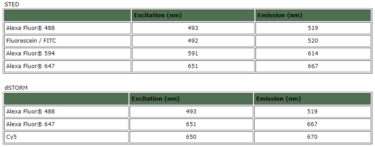
Jackson ImmunoResearch offers secondary antibodies conjugated to dyes over a broad spectral range that have been successfully employed in STED: Alexa Fluor® 488, FITC, Alexa Fluor® 594 and Alexa Fluor® 647.

Stochastic Optical Reconstruction Microscopy (STORM)

Single molecule localization can be achieved using Stochastic Optical Reconstruction Microscopy (STORM) using photoswitchable fluorophores to generate images with resolution superior to those collected using conventional methods. There are a number of SRM techniques which exploit the principle of reversible saturable optically linear fluorescence (RESOLFT), using photoswitching or photoactivation of fluorescent dyes. Examples include direct stochastic optical reconstruction microscopy (dSTORM), photoactivated localization microscopy (PALM), fluorescence photoactivation localization microscopy (fPALM), ground state depletion followed by individual molecule return (GSDIM), and super-resolution optical fluctuation imaging (SOFI).
These techniques can be performed using either a pair of dyes to function as an effector and an activator dye to achieve photoswitching, or using dyes which “self-switch” in the case of direct STORM (dSTORM) (Heilemann et al., 2008). Jackson ImmunoResearch provides secondary antibody conjugates suitable for dSTORM applications. Briefly, a relatively low-intensity excitation laser excites the sample, randomly activating a small number of dye molecules. The individually fluorescing dye molecules are spread apart enough so the center of their point spread function can be calculated, which infers the exact location of the dye. A second laser is then used to switch all the molecules off and the process is repeated. High resolution images are then mathematically generated by overlapping all of the mapped point spread functions of each dye.

Fluorophore-conjugated antibodies for single molecule localization experiments

The best dyes for single molecule localization are typically very bright and result in enough photons to reliably produce tight Gaussian distributions. Jackson ImmunoResearch offers several proven dyes in a broad spectral range such as Alexa Fluor® 488, Alexa Fluor® 647 and Cy™5 for use in these types of experiments.

Suggested fluorescent dye conjugates for super-resolution microscopy
Reference:
Huang, B., Bates, M., & Zhuang, X. (2009). Super-resolution fluorescence microscopy.
Annual Review of Biochemistry, 78 , 993–1016.
Farahani, J.N., et al. (2010). Stimulated Emission Depletion (STED) Microscopy: from Theory to Practice.
Microscopy: Science, Technology, Applications and Education. 1539-1547.
Heilemann M,et al., (2008) Subdiffraction-resolution fluorescence imaging with conventional fluorescent probes. Angew Chem Int Ed Engl. 47:6172–6176
Dempsey, et al. (2011). Evaluation of fluorophores for optimal performance in localization-based super-resolution imaging. Nature Methods, 8, 1027-1036.

For further questions CONTACT US

anti-Giardia lamblia antibody IgM, IgG and IgA ELISA Kit

Giardia lamblia (also known as Giardia intestinalis) has a characteristic tear-drops hape and measures 1015 µm in length. Giardia has a two stage life cycle consisting of trophozoite and cyst. The life cycle begins with ingested cysts, which release trophozoites (1020m x 515m) in the duodenum. These trophozoites attach to the surface of the intestinal epithelium using a ventral sucking disk and then reproduce by binary fission. The trigger for encystment is unclear, but the process results in the inactive, environmentally resistant form of Giardia cyst (1114m x 710m) that is excreted in feces.


This microplate based ELISA (enzyme linked immunosorbent assay) kit is intended for the qualitative detection of anti Giardia  labmlia IgM, IgG or IgA antibody in test sample. The assay is a useful tool in the aid of determination of Giardia  lamblia infection in acute or chronic gastroenteritis.

The assay utilizes the microplate based enzyme immunoassay technique by coating highly purified and inactive Giardia  lamblia antigen on to the wall of microtiter plate.

Human Anti-Giardia lamblia IgG ELISA Kit

Human Anti Giardia lamblia IgM ELISA Kit

Human Anti Giardia lamblia IgA ELISA Kit

For further questions CONTACT US

Posted in Uncategorized

New distribution ASSAYGATE

ELISA and ultra-sensitive immunoassay products for preclinical research, ultra-sensitive assay kits established upon Simoa (Single Molecule Array) technology platform to detect targets difficult or impossible to measure with conventional assays.

AssayGate are one of the preferred partners of Quanterix. The digital nature of the Simoa technology allows an average of 1000× sensitivity increase over conventional assays with CVs <10%. These assay research areas focus on protein biomarkers for tumors, angiogenesis, inflammation, cardiovascular diseases, metabolism, endocrinology, immunology, immunotoxicity and acute phase reactants in species including human, mouse and rat, etc
AssayGate provides the most cost-effective testing services and immunoassay products to customers in life science, biomedical and pharmaceutical communities.
Unlike many other immunoassay kit providers, AssayGate provide their own manufactured immunoassay kits in the US. This allows to offer unparalleled customer services and technical support.  All products are for research use only. Not for use in diagnostic or therapeutic procedures.   Human ELISA Kits:

CAT #

118931 Androgen receptor (AR), C-terminal

118930 Androgen receptor (AR), N-terminal

118909 AXL

118910 Bone alkaline phosphatase (BALP)

118907 Cytidine deaminase (CDA)

118920 c-Met

118937 Enolase 1 or Alpha-enolase (ENO1)

118906 Enolase 3 (ENO3)

118911 Growth Arrest-Specific 6 (GAS6)

118932 Hemoglobin (Hb)

118933 Hemoglobin A1c (HbA1c)

118913 Insulin-like growth factor 1 receptor (IGFIR)

118914 Lipopolysaccharide binding protein (LBP)

118916 Lactate dehydrogenase -5 (LDH-5)

118912 Lactate dehydrogenase A (LDHA)

118904 Midkine

118917 Mucin 4 (MUC-4)

118903 Neuregulin 1- β1 (NRG1- β1)

118927 Programmed cell death protein 1 (PD-1)

118928 Programmed death-ligand 1 (PD-L1)

118929 Programmed death-ligand 2 (PD-L2)

118918 Regenerating islet-derived family, member 4 (REG4)

118934 S100A8

118935 S100A9

118915 Semaphorin 3C (SEMA3C)

118936 Transforming growth factor beta receptor III (TGFBR3)

118905 Tyrosine kinase with immunoglobulin-like and EGF-like domains 1 (TIE-1)

118919 UL16 binding protein 2 (ULBP2)

118902 Vascular endothelial growth factor 121 (VEGF-121)

118901 von Willebrand Factor (vWF)

If you don’t see exactly what you’re looking for, contact us !

Posted in Uncategorized

DenHyb Hybridization Solutions

FULL RANGE OF PRODUCTS Click here

DESCRIPTION:
Hybridization Solution to dilute probes for a better efficiency of FISH and ISH
BENEFITS:

Reduce costs with a dilution of the probe 5 to 40 times

Saving probes  Compatible with a wide range of probes

Suitable for most protocols

Stable for more than a year at -20 ° C

Two types of DenHyb: for cell and tissue

PROTOCOL: denhyb_protocol

LITERATURE: denhyb_citations

Posted in Uncategorized

Myostatin ELISA/EIA Kit

INTENDED USE

This enzyme immunoassay is intended for the quantitative determination of myostatin in human serum and plasma. For in vitro diagnostic use only.

SUMMARY

Myostatin belongs to the transforming growth differentiation factor-ß (TGF-ß) super family. The molecule is a negative regulator of muscle growth, but details about the actions of myostatin are uncertain (Roth and Walsh, 2004).

Myostatin was first identified in 1997 by McPherron et al. They found out that null-mutant knockout mice were significantly larger than wild-type animals and exhibited a large and widespread increase in skeletal muscle mass due to an increase of muscle fiber number (hyperplasia) and thickness (hypertrophy). Other groups identified mutations in the myostatin gene in naturally bred “double-muscles” cattle breeds.


Similar to the findings in animal models, increased myostatin immuno-reactivity or expression has been observed in HIV-infected men with muscle wasting (Gonzales-Cadavid et al. 1998), after prolonged bed rest in young men (Zachwieja et al. 1999) and in older men and women with muscle wasting (Yarasheski KE et al. 2002).


Shi et al. (2007) and others have found that myostatin deficiency inhibits adipogenesis in vivo, even when mice are fed a high-fat diet. Transgenic overexpression of myostatin pro-peptide, which inhibits myostatin signaling, also inhibits body fat gain with a high-fat diet (Zhao et al. 2005). Similar alterations in myostatin signaling are associated with changes in body fat among humans.

INDICATIONS

  • Regulation of muscle growth
  • Muscle atrophy
  • Muscle wasting

Product: IMM-K1012 Myostatin ELISA/EIA Kit

For further questions CONTACT US

Posted in Uncategorized

GLP-1 ELISA Kit

INTENDED USE

This high sensitive ELISA (enzyme-linked immunosorbent assay) kit is produced for the exclusively quantitative determination of glucagon-llike peptide-1 (7-36) [GLP-1 (7-36)] level in serum and EDTA-plasma sample among human, rat, mouse, goat, etc.


ASSAY PRINCIPLE

This ELISA is designed, developed and produced for the quantitative measurement of bioactive GLP-1 (7-36) in plasma sample. The assay utilizes the two-site “sandwich” technique with two selected GLP-1 (7-36) specific antibodies. Assay standards, controls and test samples are directly added to wells of a microplate that is coated with streptavidin. Subsequently, a mixture of biotinylated GLP-1 (7-36) specific antibody and a horseradish peroxidate (HRP) conjugated GLP-1 (7-36) specific antibody is added to each well. After the first incubation period, a “sandwich” immunocomplex of “Streptavidin – Biotin-Antibody – GLP-1(7-36) – HRP conjugated antibody” is formed and attached to the wall of the plate. The unbound HRP conjugated antibody is removed in a subsequent washing step. For the detection of this immunocomplex, each well is then incubated with a substrate solution in a timed reaction and then measured in a spectrophotometric microplate reader. The enzymatic activity of the immunocomplex bound to GLP-1 (7-36) on the wall of the microtiter well is directly proportional to the amount of  GLP-1 (7-36) in the sample.


Active GLP-1 (7-36) ELISA

Measures GLP-1 (7-36) without cross reaction to:

– GLP-1 (9-36), (7-37), (9-37), (1-36), (1-37)

– GLP-2

– Glucagon

Assay Sensivity: 0.6 pmol/l


Total GLP-1 ELISA

Measures the sum value of GLP-1 (7-36) and (9-36) without cross reaction to:

– GLP-1 (7-37), (9-37), (1-36), (1-37)

– GLP-2

– Glucagon

Assay Sensivity: 1.0 pmol/l

PRODOTTI:

Active GLP-1 (7-36) ELISA Cat # EPI-KT871

Total GLP-1 ELISA Cat # EPI-KT876

GLP-1 Sample Extraction Kit Cat # EPI-KT910

For further questions CONTACT US